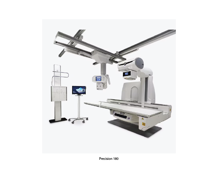
Precision 180
The Precision 180 Radiography and Fluoroscopy System is an advanced medical imaging system designed to provide high-quality radiographic and fluoroscopic images. This system is used for a wide range of diagnostic applications, offering both versatility and precision in imaging. Here are some of its key features and benefits.
Key Features of Precision 180:
1. Versatile Imaging Capabilities:
• Radiography and Fluoroscopy: Combines both radiographic and fluoroscopic functionalities in one system, making it suitable for a variety of diagnostic procedures.
• Dynamic Imaging: Capable of capturing dynamic processes in real-time, which is essential for fluoroscopic examinations.
2. Advanced Imaging Technology:
• High-Resolution Detector: Provides high-quality, detailed images with excellent contrast resolution.
• Low Dose Imaging: Utilizes advanced technology to minimize radiation exposure to patients while maintaining image quality.
3. User-Friendly Design:
• Ergonomic Interface: Designed for ease of use, with an intuitive interface that allows technologists to perform exams efficiently.
• Automated Positioning: Features automated positioning tools that streamline the workflow and reduce the time required for imaging procedures.
4. Patient Comfort and Safety:
• Flexible Table Design: The table design accommodates various patient positions and body types, enhancing patient comfort during exams.
• Safety Features: Includes various safety mechanisms to protect patients and operators from unnecessary radiation exposure.
5. Enhanced Diagnostic Capabilities:
• Digital Subtraction Angiography (DSA): Available in some models, this feature helps visualize blood vessels by subtracting bone and soft tissue structures from the image.
• Image Processing Tools: Advanced software tools for image processing and enhancement improve diagnostic accuracy.
Key Features of Precision 180:
1. Interventional Radiology:
• Angiography: Used for diagnosing and treating vascular diseases by visualizing blood vessels and guiding catheter-based interventions such as angioplasty and stent placement.
• Embolization: Assists in procedures to block blood flow to abnormal tissues, such as tumors or aneurysms, by guiding the delivery of embolic agents.
• Biopsies and Drainages: Provides real-time imaging guidance for precise needle placement in biopsies and fluid drainages.
2. Interventional Cardiology:
• Coronary Angiography: Visualizes coronary arteries to diagnose blockages and guide interventions such as angioplasty and stenting.
• Electrophysiology (EP) Procedures: Assists in mapping and ablating abnormal cardiac rhythms by providing detailed images of cardiac anatomy and catheter positions.
• Structural Heart Interventions: Supports procedures such as transcatheter aortic valve replacement (TAVR) and mitral valve repair by offering precise imaging and guidance.
3. Oncology:
• Tumor Ablation: Guides the placement of ablation probes for thermal or chemical destruction of tumors.
• Targeted Drug Delivery: Assists in the precise delivery of chemotherapy or other therapeutic agents directly to tumors, minimizing systemic exposure and side effects.
4. Neurointerventions:
• Cerebral Angiography: Visualizes cerebral vasculature to diagnose and treat conditions such as aneurysms, arteriovenous malformations (AVMs), and stroke.
• Endovascular Therapy: Guides interventions such as coiling of aneurysms or mechanical thrombectomy in stroke patients.
5. Spinal and Musculoskeletal Interventions:
• Vertebroplasty/Kyphoplasty: Assists in the injection of bone cement to stabilize compression fractures in the spine.
• Pain Management: Guides the placement of needles for epidural steroid injections or nerve blocks.
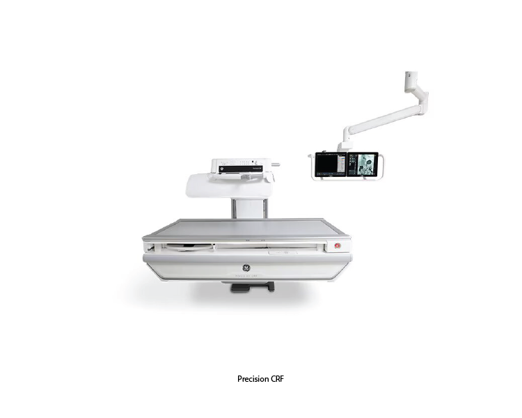
Precision CRF
The Precision CRF (Compact Radiography and Fluoroscopy) system is an advanced imaging solution designed for a wide range of diagnostic applications. It combines radiographic and fluoroscopic capabilities into a single, compact unit, offering high-quality imaging with enhanced patient comfort and operational efficiency. Here are some key features and benefits of the Precision™ CRF system.
Key Features of Precision CRF:
1. Integrated Radiography and Fluoroscopy:
• Dual Functionality: Combines radiography and fluoroscopy in one system, allowing for versatile imaging applications from static X-rays to dynamic studies.
• Seamless Transition: Smooth transition between radiographic and fluoroscopic modes to streamline workflow and improve efficiency.
2. High-Quality Imaging:
• Advanced Detectors: Equipped with high-resolution digital detectors that provide clear and detailed images with excellent contrast.
• Dose Efficiency: Utilizes advanced technology to minimize radiation dose while maintaining superior image quality, ensuring patient safety.
3. Compact and Ergonomic Design:
• Space-Saving: Compact design ideal for facilities with limited space without compromising functionality.
• Ergonomics: User-friendly design with easy-to-use controls and interfaces to enhance operator comfort and efficiency.
4. Enhanced Patient Comfort:
• Adjustable Table: Features a patient table that can be adjusted to accommodate various patient sizes and conditions, ensuring comfort during procedures.
• Gentle Positioning: Smooth and gentle movements reduce patient discomfort and anxiety.
5. Versatile Clinical Applications:
• Wide Range of Exams: Suitable for a variety of diagnostic exams including gastrointestinal studies, orthopedic assessments, and vascular imaging.
• Real-Time Imaging: Fluoroscopy capabilities provide real-time imaging for dynamic studies such as swallowing assessments and catheter placements.
6. Advanced Imaging Technology:
• Digital Subtraction Angiography (DSA): Some models include DSA capabilities to enhance vascular imaging by removing bone and tissue from the image.
• Image Processing: Advanced image processing tools enhance diagnostic accuracy by improving image clarity and detail.
Clinical Applications of Precision CRF
1. Oncology:
• Liver Tumors: Effective for treating primary liver cancers (hepatocellular carcinoma) and metastatic liver tumors. Both cryoablation and RFA can be used to target and destroy tumor cells with minimal impact on liver function.
• Kidney Tumors: Particularly useful for small renal tumors. Cryoablation is favored for tumors near critical structures due to its controlled ice ball formation.
• Lung Tumors: Offers a minimally invasive option for patients with primary or metastatic lung cancer who are not candidates for surgery.
2. Bone and Soft Tissue Tumors:
• Osteoid Osteoma: RFA is commonly used to treat this benign but painful bone tumor. The precision of the system ensures effective treatment while preserving bone integrity.
• Metastatic Bone Disease: Provides palliative treatment to reduce pain and stabilize bones affected by metastatic cancer.
3. Pain Management:
• Nerve Ablation: Can be used to ablate nerves responsible for chronic pain, providing significant pain relief for patients with conditions like chronic lower back pain or arthritis.
4. Prostate Cancer:
• Focal Therapy: Cryoablation offers a minimally invasive option for focal therapy in prostate cancer, targeting only the cancerous tissue while sparing the rest of the prostate and reducing side effects.
5. Benign Tumors and Other Conditions:
• Fibroadenomas: Cryoablation can be used to treat benign breast tumors, offering a less invasive alternative to surgical removal.
• Benign Prostatic Hyperplasia (BPH): RFA can be used to shrink the prostate and relieve symptoms of BPH.