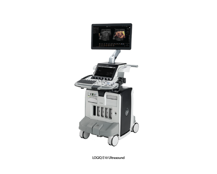
LOGIQ E10
The LOGIQ E10 ultrasound system by GE Healthcare is a state-of-the-art imaging tool designed to meet the needs of diverse clinical environments. With its advanced imaging technologies, AI-driven automation, and ergonomic design, it provides superior image quality, enhances workflow efficiency, and supports a wide range of clinical applications.
Key Features of LOGIQ E10 Ultrasound
1. cSound™ Architecture:
• Utilizes a next-generation platform with cSound™ Imageformer, which provides powerful parallel processing and high-resolution imaging, delivering exceptional image quality.
2. Advanced Imaging Technologies:
• XDclear™ Probes: High-performance transducers that offer deep penetration and high resolution, improving diagnostic confidence across various patient body types.
• Photo Assistant: Integrates photos of anatomical landmarks with ultrasound images for better documentation and reporting.
• Automated Functionality: Includes features such as auto-optimized images, automated measurement tools, and 3D/4D imaging capabilities to enhance efficiency and ease of use.
3. Artificial Intelligence (AI) and Automation:
• AI-driven tools and algorithms, such as Auto Lesion Segmentation and Auto IMT (Intima-Media Thickness), to streamline workflows and improve diagnostic accuracy.
• Scan Assistant: Customizable protocols that guide the user through exams, improving consistency and reducing scan times.
4. Ergonomic Design:
• User-friendly interface with customizable touchscreen controls, adjustable monitor, and ergonomic transducer design to enhance user comfort and reduce strain during prolonged use.
5. Comprehensive Connectivity:
• Seamless integration with hospital information systems (HIS), picture archiving and communication systems (PACS), and other digital platforms for efficient data management and sharing.
6. High-Resolution Imaging:
• Exceptional image clarity and detail across a wide range of clinical applications, thanks to advanced beam forming and sophisticated image processing technologies.
Clinical Applications of LOGIC E10
1. Abdominal Imaging:• High-resolution imaging for detailed assessment of abdominal organs, including the liver, kidneys, pancreas, and gallbladder.
2. Cardiology:
• Comprehensive cardiac imaging capabilities, including echocardiography for evaluating heart structure and function.
3. Obstetrics and Gynecology:
• Advanced 3D/4D imaging for fetal assessment, as well as detailed evaluation of gynecological structures.
4. Musculoskeletal Imaging:
• High-resolution imaging of muscles, tendons, ligaments, and joints for diagnosing injuries and conditions affecting the musculoskeletal system.
5. Vascular Imaging:
• Detailed visualization of blood vessels for assessing vascular conditions, including arterial and venous diseases.
6. Pediatrics:
• Tailored imaging protocols and high-resolution capabilities for evaluating pediatric patients, ensuring accurate and gentle assessments.
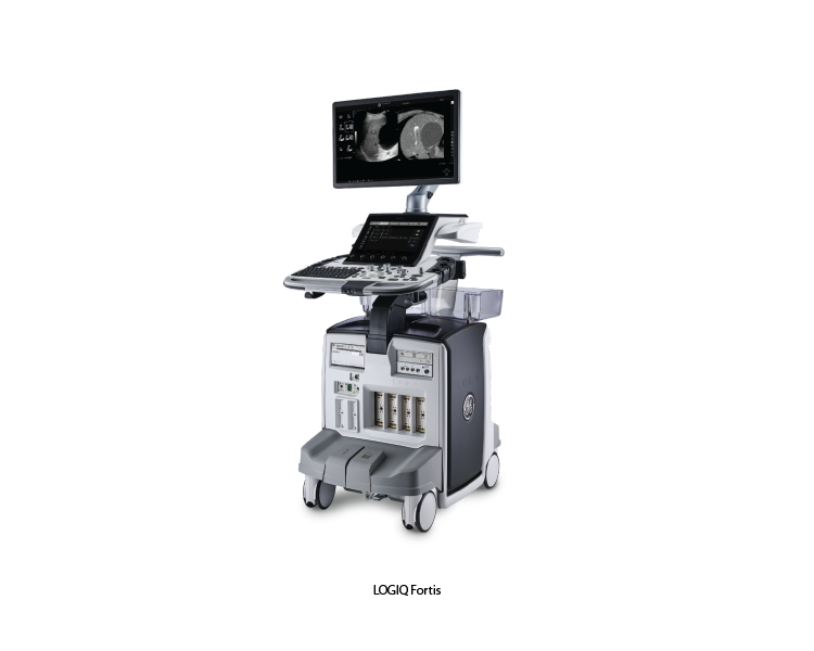
LOGIQ Fortis
The LOGIQ Fortis Ultrasound system, developed by GE Healthcare, represents a new standard in general imaging ultrasound. It is designed to provide exceptional image quality, advanced clinical capabilities, and streamlined workflows. With its versatile and robust design, the LOGIQ Fortis is suitable for a wide range of clinical applications, from routine exams to complex diagnostic procedures. This system aims to enhance diagnostic confidence and improve patient care through its innovative features and user-centric design.
Key Features of LOGIC Fortis
1. Superior Imaging Quality:
• High-Resolution Imaging: Equipped with advanced transducers and imaging technologies that deliver clear, detailed images with high resolution and contrast.
• Advanced Imaging Modes: Includes harmonic imaging, speckle reduction imaging, and 3D/4D imaging for comprehensive and precise diagnostic assessments.
2. Advanced Clinical Tools:
• Elastography: Provides strain and shear wave elastography for assessing tissue stiffness, aiding in the diagnosis of liver disease, breast lesions, and other conditions.
• Contrast-Enhanced Ultrasound (CEUS): Enhances the visualization of blood flow and tissue perfusion using ultrasound contrast agents, useful for liver and vascular imaging.
• Auto-Optimization: Features automated tools for optimizing image quality, such as Auto Image Optimization and Auto Doppler, to enhance workflow efficiency.
3. User-Friendly Interface:
• Intuitive Touchscreen: Large, high-resolution touchscreen interface for easy navigation and quick adjustments during exams.
• Customizable Settings: Allows users to personalize presets and protocols to match specific workflow preferences and clinical requirements.
4. Ergonomic and Portable Design:
• Compact and Mobile: Designed with a small footprint and wheels for easy mobility within the clinical environment, facilitating bedside and point-of-care imaging.
• Ergonomic Features: Adjustable control panel and monitor to enhance user comfort and reduce strain during extended use.
5. Comprehensive Connectivity:
• DICOM Compatibility: Ensures seamless integration with hospital information systems (HIS) and picture archiving and communication systems (PACS), facilitating efficient data management.
• Wireless Capabilities: Supports wireless data transmission for easy image sharing and integration within clinical networks.
Clinical Applications of LOGIC Fortis
1. General Imaging:
• Provides high-quality imaging for a wide range of applications including abdominal, pelvic, thyroid, breast, and small parts examinations.
2. Vascular Imaging:
• Advanced Doppler and CEUS capabilities allow for detailed vascular assessments, including evaluation of blood flow and detection of vascular abnormalities.
3. Musculoskeletal Imaging:
• High-resolution imaging of muscles, tendons, and joints to assist in diagnosing injuries and conditions related to the musculoskeletal system.
4. Obstetrics and Gynecology:
• Offers detailed fetal and maternal imaging, aiding in prenatal care, fetal monitoring, and gynecological assessments.
5. Elastography Applications:
• Used for assessing tissue stiffness in various clinical settings, including liver fibrosis evaluation, breast lesion characterization, and thyroid nodule assessment.